Knee Segmentation Model Development Using Swin-Unet
📌 Overview
This project implements a Swin UNet model for automatic segmentation of the knee joint from MRI scans, followed by a 3D reconstruction of anatomical structures. Swin UNet leverages the power of transformer-based architectures for improved contextual understanding in medical image segmentation.
🏗️ Swin UNet Architecture
Swin UNet is based on the Swin Transformer backbone, which applies shifted window attention to efficiently model long-range dependencies in the image. It maintains a hierarchical representation and integrates it into a UNet-like encoder-decoder design.
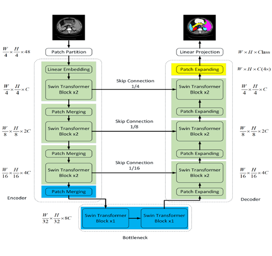
Figure: Swin UNet Architecture
🧠 3D Reconstruction Results
After applying Swin UNet for automatic segmentation of the knee from MRI scans, we performed a 3D reconstruction of the segmented anatomical parts. The video below showcases the segmented femur and tibia rendered in 3D for better visual analysis.
Video: 3D Reconstruction of the Segmented Knee
🔧 Tools & Technologies
- Python
- PyTorch
- MONAI (Medical Open Network for AI)
- VTK & Matplotlib for 3D Visualization
📝 Conclusion
This project demonstrates the application of transformer-based architectures in medical imaging. Swin UNet offers powerful performance for anatomical segmentation, while 3D reconstruction allows for more intuitive interpretation of medical scans.